Gynecomastia Returning on One Side: How to Tell If It Is a Recurrence
Author : Dr. Young-jin SimMay 7, 2026 at 10:53 AM 179
Gynecomastia Returning on One Side:
How to Tell If It Is a Recurrence
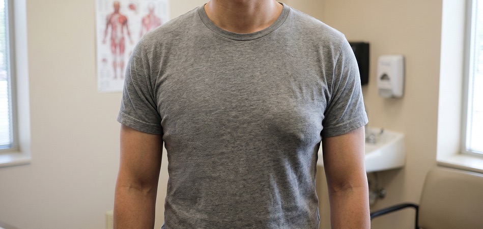
1. Why Only One Side Changes
2. Distinguishing True Recurrence, Fat Re-accumulation, and Residual Tissue
| Category | True recurrence | Fat re-accumulation | Residual tissue |
|---|---|---|---|
| Cause | Remaining glandular tissue regrows under hormonal stimulation |
Chest fat re-accumulates with weight gain |
Glandular tissue not removed in the initial surgery remains |
| Feel | Firm, well-defined lump beneath the nipple |
Soft, poorly defined fatty feel |
Thin disc-shaped resistance around the nipple |
| Timing | Progresses gradually from 6 months to several years after surgery |
Appears in proportion to weight changes |
Palpable continuously from right after surgery |
| Ultrasound findings | Reappearance of hypoechoic glandular tissue shadow |
Only uniform fat echoes observed |
Small amount of residual glandular tissue confirmed just below the nipple |
| Response | Hormone testing plus consideration of additional gland excision |
Weight management first, liposuction if needed |
Observation or decision on additional excision |
Steps to Identify a One-Sided Change
How to Confirm Whether It Is a Recurrence
If you feel that one side has come back,
rather than worrying alone, get both an ultrasound and a hormone test.
Depending on the cause, observation may be sufficient, or further measures may be needed.
An accurate diagnosis is the fastest way to reduce unnecessary worry.
References
- Gynecomastia review (etiology, mechanism, course) — Braunstein GD. Gynecomastia. N Engl J Med. 2007;357(12):1229–1237. doi:10.1056/NEJMcp070677
- Drug-induced gynecomastia — Bowman JD, et al. Drug-induced gynecomastia. Pharmacotherapy. 2012;32(12):1123–1140. doi:10.1002/phar.1138
Read next — Doctor Column
All Doctor Columns →Customer Story Column
All Customer Story Columns →
Key Background
- · Board Certified Urologist / M.S. in Medicine
- · M.D., Kyungpook National University School of Medicine
View Full Profile ›
"A doctor proven by results and remembered for sincerity — a finish where the detail is different."
"Surgery is a matter of technique, but caring for a patient is always a matter of the heart. I explain not only the result but the process honestly, and think it through together with the patient so they can choose after fully understanding. Through years of male body contouring and men's surgery, one standard has not changed: to take responsibility from before the operation through afterwards, so that the patient does not regret this choice. That, to me, is what medical care is."
- · Residency, Samsung Medical Center, Seoul
- · Fellowship, Gachon University Gil Hospital
- · Member, Korean Society for Sexual Medicine and Andrology (KSSMA)
- · Member, Korean Academy of Aesthetic Medicine
- · Member, Korean Prostate Society (KPS)
- · Member, Korean Association of Breast Surgery (KABS)
- · Member, Union Internationale de Médecine Esthétique (U.I.M.E.)
- · Former Director, Trueman Men's Clinic Gangnam
- · Member, Korean Association of Urogenital Tract Infection and Inflammation (KAUTII) / Korean Society of Urological Infection